
Carotid Duplex Ultrasound
A non-invasive ultrasound examination of the carotid arteries in the neck. Findings are interpreted together with the clinical question and referring clinician's assessment.
New stroke-like symptoms are an emergency
Sudden face drooping, weakness or numbness on one side, speech difficulty, vision loss, or severe new imbalance needs emergency care even if it improves or disappears. Call 112 or go to the nearest emergency department immediately; do not book a routine scan or wait for WhatsApp.
The booking option below is for stable, clinician-directed carotid assessment or follow-up only; this page cannot diagnose a stroke.
Expected duration
20-40 minutes
Preparation
Usually no special preparation
Report
As arranged
Comfort
Usually well tolerated
What is Carotid Duplex Ultrasound?
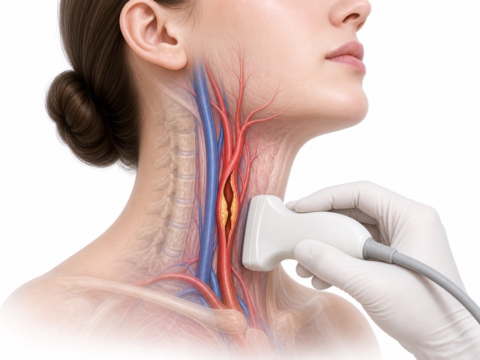
Carotid duplex ultrasound uses sound waves to examine the carotid arteries in the neck and assess the direction and speed of blood flow.
It may identify plaque, narrowing, or other blood-flow changes. Results must be interpreted with symptoms, history, and the treating clinician's assessment.
Common Reasons for Referral
- Previous stroke or transient ischemic attack (TIA) when carotid imaging is requested by a clinician
- A clinician-defined carotid imaging question after urgent assessment of new focal neurological symptoms, such as sudden weakness, numbness, speech difficulty, or temporary vision loss
- A neck bruit found during examination as part of a clinician-defined work-up
- Follow-up of known carotid plaque, narrowing, or established carotid disease
- Surveillance before or after a selected carotid procedure when requested by the treating team
- A specific referral question identified by the treating clinician; vascular risk factors are assessed in context and do not by themselves automatically require routine scanning
What Happens During the Examination
- 1You lie comfortably with the neck positioned for the examination
- 2Ultrasound gel is applied to the neck
- 3The transducer is moved gently along both sides of the neck
- 4Images and blood-flow measurements are recorded
- 5The findings are documented for clinical review
Frequently Asked Questions
Related Vascular Services
Explore our comprehensive vascular diagnostic services available at International Hospital, Kuwait